Beranda
/ Anatomy Of Chest : Human Chest Anatomy Illustration Stock Image F025 1027 Science Photo Library / It's also sometimes referred to as the breastbone.
Anatomy Of Chest : Human Chest Anatomy Illustration Stock Image F025 1027 Science Photo Library / It's also sometimes referred to as the breastbone.
Insurance Gas/Electricity Loans Mortgage Attorney Lawyer Donate Conference Call Degree Credit Treatment Software Classes Recovery Trading Rehab Hosting Transfer Cord Blood Claim compensation mesothelioma mesothelioma attorney Houston car accident lawyer moreno valley can you sue a doctor for wrong diagnosis doctorate in security top online doctoral programs in business educational leadership doctoral programs online car accident doctor atlanta car accident doctor atlanta accident attorney rancho Cucamonga truck accident attorney san Antonio ONLINE BUSINESS DEGREE PROGRAMS ACCREDITED online accredited psychology degree masters degree in human resources online public administration masters degree online bitcoin merchant account bitcoin merchant services compare car insurance auto insurance troy mi seo explanation digital marketing degree floridaseo company fitness showrooms stamfordct how to work more efficiently seowordpress tips meaning of seo what is an seo what does an seo do what seo stands for best seotips google seo advice seo steps, The secure cloud-based platform for smart service delivery. Safelink is used by legal, professional and financial services to protect sensitive information, accelerate business processes and increase productivity. Use Safelink to collaborate securely with clients, colleagues and external parties. Safelink has a menu of workspace types with advanced features for dispute resolution, running deals and customised client portal creation. All data is encrypted (at rest and in transit and you retain your own encryption keys. Our titan security framework ensures your data is secure and you even have the option to choose your own data location from Channel Islands, London (UK), Dublin (EU), Australia.
Anatomy Of Chest : Human Chest Anatomy Illustration Stock Image F025 1027 Science Photo Library / It's also sometimes referred to as the breastbone.. The thorax has two major openings: It provides access to ct images in the axial plane, allowing the user to learn and review the lung anatomy interactively. The chest or thorax region of the upper body has a number of important organs that reside within it that may present with chest pain if they become compromised in. The epidermis is the outermost layer that provides a protective, waterproof seal over the body. Organs & structures of the chest heart.
The dominant muscle in the upper chest is the pectoralis major. Organs & structures of the chest heart. The superior thoracic aperture found superiorly and the inferior thoracic aperture. The chest or thorax region of the upper body has a number of important organs that reside within it that may present with chest pain if they become compromised in. In insects, crustaceans, and the extinct trilobites, the thorax is one of the three main divisions of the creature's body, each of which is in turn composed of multiple segments.
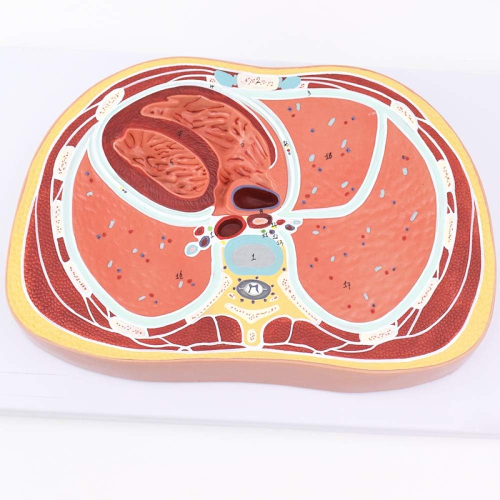
Human Thoracic Transection Model Cardiopulmonal Model Human Cross Section Anatomy Chest Horizontal Cross Section Model Amazon De Kuche Haushalt from images-na.ssl-images-amazon.com In insects, crustaceans, and the extinct trilobites, the thorax is one of the three main divisions of the creature's body, each of which is in turn composed of multiple segments. The thorax has two major openings: Browse 2,509 female chest anatomy stock photos and images available, or start a new search to explore more stock photos and images. The major muscle in the chest is the pectoralis major. It spreads out like a fan and covers the rib cage like an armor plate. Chest a man's chest — like the rest of his body — is covered with skin that has two layers. The circulatory system does most of its work. Anatomy of the chest and the lungs:
Anatomy of the chest, abdomen, and pelvis was produced in part due to the generous funding of the david f.
The chest or thorax region of the upper body has a number of important organs that reside within it that may present with chest pain if they become compromised in. The first rib is a short, flat rib that is much wider and more curved than those previously described. Thoracic cavity, also called chest cavity, the second largest hollow space of the body. The chest is the area of origin for many of the body's systems as it houses organs such as the heart, esophagus, trachea, lungs, and thoracic diaphragm. The chest anatomy includes the pectoralis major, pectoralis minor and the serratus anterior. Swensen fund for innovation in teaching. Chest muscles anatomy (1) pectoralis major muscle. Learn about each of these muscles, their locations, functional anatomy and exercises for them. (1) the pectoralis major, and (2) the pectoralis minor. It spreads out like a fan and covers the rib cage like an armor plate. Structures to identify • heart • lungs • mediastinum • pleural space • chest wall • …everything else! The dominant muscle in the upper chest is the pectoralis major. Radiology basics of chest ct anatomy with annotated coronal images and scrollable axial images to help medical students and junior doctors learning anatomy.
Anatomy of the thorax, heart, abdomen and pelvis recommended text gray's anatomy for students. Computed tomography (ct) of the chest can detect pathology that may not show up on a conventional chest radiograph (1). Organs & structures of the chest heart. (1) the pectoralis major, and (2) the pectoralis minor. The circulatory system does most of its work.
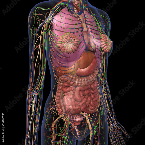
Female Anatomy Of Chest And Abdomen On Black Background 3 Buy This Stock Photo And Explore Similar Images At Adobe Stock Adobe Stock from as2.ftcdn.net This thoracic and pulmonary anatomy tool is especially designed for students of anatomy (medical and paramedical studies). Your sternum is a bone that's located in the middle of your chest. The chest or thorax region of the upper body has a number of important organs that reside within it that may present with chest pain if they become compromised in. The human thorax includes the thoracic cavity and the thoracic wall. The chest wall is comprised of skin, fat, muscles, and the thoracic skeleton. It provides protection to vital organs (eg, heart and major vessels, lungs, liver) and provides stability for movement. It is generally agreed that radiographic examination of the chest is extremely important in the diagnosis of pulmonary disease. It is enclosed by the ribs, the vertebral column, and the sternum, or breastbone, and is separated from the abdominal cavity (the body's largest hollow space) by a muscular and membranous partition, the diaphragm.
The chest is the area of origin for many of the body's systems as it houses organs such as the heart, esophagus, trachea, lungs, and thoracic diaphragm.
Anatomy of the chest and shoulder, anatomy of the chest organs, anatomy of the chest wall, anatomy of the chest wall and pleura, anatomy of upper chest area, human. The chest or thorax region of the upper body has a number of important organs that reside within it that may present with chest pain if they become compromised in. The circulatory system does most of its work. The major muscle in the chest is the pectoralis major. Learn about each of these muscles, their locations, functional anatomy and exercises for them. The human thorax includes the thoracic cavity and the thoracic wall. The thorax has two major openings: Thoracic cavity, also called chest cavity, the second largest hollow space of the body. Choose from 500 different sets of anatomy chest flashcards on quizlet. The first step in understanding thorax anatomy is to find out its boundaries. Your sternum protects the organs of your torso from injury and also serves as a. Anatomy of the chest and the lungs: The superior thoracic aperture found superiorly and the inferior thoracic aperture.
Chest muscles anatomy (1) pectoralis major muscle. The thorax has two major openings: Learn about each of these muscles, their locations, functional anatomy and exercises for them. It's also sometimes referred to as the breastbone. The circulatory system does most of its work.
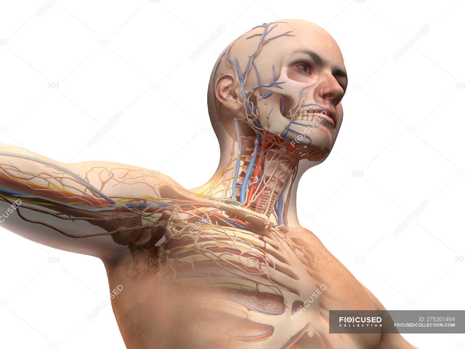
Male Head And Chest Anatomy Diagram With Ghost Effect On White Background 3d Lungs Stock Photo 275201494 from st.focusedcollection.com The superior thoracic aperture found superiorly and the inferior thoracic aperture. Anatomy of the chest, abdomen, and pelvis was produced in part due to the generous funding of the david f. It spreads out like a fan and covers the rib cage like an armor plate. (1) the pectoralis major, and (2) the pectoralis minor. Learn anatomy chest with free interactive flashcards. Radiology basics of chest ct anatomy with annotated coronal images and scrollable axial images to help medical students and junior doctors learning anatomy. Anatomy of the chest and shoulder, anatomy of the chest organs, anatomy of the chest wall, anatomy of the chest wall and pleura, anatomy of upper chest area, human. This page provides an overview of the chest muscle group.
Chest muscles anatomy (1) pectoralis major muscle.
Your sternum protects the organs of your torso from injury and also serves as a. This page provides an overview of the chest muscle group. Thoracic cavity, also called chest cavity, the second largest hollow space of the body. It provides access to ct images in the axial plane, allowing the user to learn and review the lung anatomy interactively. It spreads out like a fan and covers the rib cage like an armor plate. In insects, crustaceans, and the extinct trilobites, the thorax is one of the three main divisions of the creature's body, each of which is in turn composed of multiple segments. The chest or thorax is the region between the neck and diaphragm that encloses organs, such as the heart, lungs, esophagus, trachea, and thoracic diaphragm. Anatomy of the chest and shoulder, anatomy of the chest organs, anatomy of the chest wall, anatomy of the chest wall and pleura, anatomy of upper chest area, human. The dominant muscle in the upper chest is the pectoralis major. Anatomy of the chest and the lungs: The epidermis is the outermost layer that provides a protective, waterproof seal over the body. Chest muscles anatomy (1) pectoralis major muscle. Swensen fund for innovation in teaching.